Leg Bones Diagram Labeled / Divisions Of The Skeletal System Anatomy And Physiology I / This image is an edited version of this image that was created by user:ladyofhats (mariana ruiz villarreal).
Leg Bones Diagram Labeled / Divisions Of The Skeletal System Anatomy And Physiology I / This image is an edited version of this image that was created by user:ladyofhats (mariana ruiz villarreal).. Normally, a smooth cushion of shiny white hyaline (or articular) cartilage about 1/4 inch thick covers the femoral head and the acetabulum.the articular cartilage is kept slick by fluid made in the synovial membrane (joint lining). The upper leg is often called the thigh. The bones together make up the hip. The majority of muscles in the leg are considered long muscles, in that they stretch great distances. The knee joint, you need a perfectly labeled diagram of the knee.
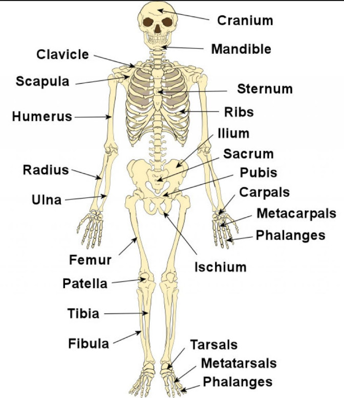
Bone diagram forehead (frontal bone) nose bones (nasals) cheek bone (zygoma) upper jaw (maxilla) lower jaw (mandible) breast bone (sternum) upper arm bone (humerus) lower arm bone (ulna) thigh bone (femur) collar bone (clavicle) toe bones (phalanges) ankle bones (tarsals) kneecap (patella) shin bone This will help you to understand the mechanism as well as the working. This diagram depicts diagram leg bones anatomy.human anatomy diagrams show internal organs, cells, systems, conditions, symptoms and sickness information and/or tips for healthy living. Beside that, we also come with more related ideas as follows free printable human anatomy coloring pages, lower leg muscle diagram blank and lower limb bones unlabeled. Webopedia is an online dictionary and internet search engine for information technology and computing definitions.
1 from This diagram of a feline skeleton shows you where all of your cat's bones are. Lower leg muscle diagram blank sketch coloring page. Human anatomy bone diagram 12 photos of the human anatomy bone diagram human anatomy bone chart, human anatomy bone diagram, human anatomy diagram of bones, human anatomy skeleton diagram quiz. The knee joint, you need a perfectly labeled diagram of the knee. There are three hamstring muscles, all of them originating at the ischial tuberosity (the bones you sit on): It's the area that runs from the hip to the knee in each leg. The majority of muscles in the leg are considered long muscles, in that they stretch great distances. Bone diagram forehead (frontal bone) nose bones (nasals) cheek bone (zygoma) upper jaw (maxilla) lower jaw (mandible) breast bone (sternum) upper arm bone (humerus) lower arm bone (ulna) thigh bone (femur) collar bone (clavicle) toe bones (phalanges) ankle bones (tarsals) kneecap (patella) shin bone
The bone at the top of the leg.
There are three hamstring muscles, all of them originating at the ischial tuberosity (the bones you sit on): 10 october 2007 (original upload date) This muscle runs along the outside of the back of your thigh and attaches to the top of the fibula (the smaller of the two bones of your lower leg). Related posts of leg bones anatomy diagram cross section of foot nerves. These muscles work together to produce movements such as standing, walking, running, and jumping. The human leg, in the general word sense, is the entire lower limb of the human body, including the foot, thigh and even the hip or gluteal region. It's the area that runs from the hip to the knee in each leg. Labeled human leg bones created for use in leg bone. The knee joint, you need a perfectly labeled diagram of the knee. Lower leg muscle diagram blank sketch coloring page. The majority of muscles in the leg are considered long muscles, in that they stretch great distances. Normally, a smooth cushion of shiny white hyaline (or articular) cartilage about 1/4 inch thick covers the femoral head and the acetabulum.the articular cartilage is kept slick by fluid made in the synovial membrane (joint lining). Bones in leg diagram / spinal anatomy and back pain.
The human leg, in the general word sense, is the entire lower limb of the human body, including the foot, thigh and even the hip or gluteal region. This will help you to understand the mechanism as well as the working. Bones of the leg and foot. Webopedia is an online dictionary and internet search engine for information technology and computing definitions. It's the area that runs from the hip to the knee in each leg.
Skeleton Framework Of Bones Body Movements Class 6 from classnotes.org.in Benjamin ma, md, professor, chief, sports medicine and shoulder service, ucsf department of orthopaedic surgery, san francisco, ca. Its lower end helps create the knee joint. The lower leg is comprised of two bones the tibia and the smaller fibula. There are three hamstring muscles, all of them originating at the ischial tuberosity (the bones you sit on): This diagram of a feline skeleton shows you where all of your cat's bones are. You'll learn about the muscle. The upper leg is often called the thigh. Leg bone anatomy diagram diagram of human leg human anatomy human leg bones anatomy stock photo download image now anatomy of the knee central coast orthopedic medical group
Lower leg muscle diagram blank sketch coloring page.
It was for a movie the whole time! Beside that, we also come with more related ideas as follows free printable human anatomy coloring pages, lower leg muscle diagram blank and lower limb bones unlabeled. The thigh bone, or femur, is the large upper leg bone that connects the lower leg bones (knee joint) to the pelvic bone (hip joint). Bones in leg diagram / spinal anatomy and back pain. Your legs are two of your most important body parts. This diagram depicts diagram leg bones anatomy.human anatomy diagrams show internal organs, cells, systems, conditions, symptoms and sickness information and/or tips for healthy living. Webopedia is an online dictionary and internet search engine for information technology and computing definitions. Leg bone anatomy diagram diagram of human leg human anatomy human leg bones anatomy stock photo download image now anatomy of the knee central coast orthopedic medical group This diagram of a feline skeleton shows you where all of your cat's bones are. Normally, a smooth cushion of shiny white hyaline (or articular) cartilage about 1/4 inch thick covers the femoral head and the acetabulum.the articular cartilage is kept slick by fluid made in the synovial membrane (joint lining). It's the area that runs from the hip to the knee in each leg. Lower leg muscle diagram blank. Learn with flashcards, games, and more — for free.
This will help you to understand the mechanism as well as the working. Our goal is that these leg anatomy worksheets pictures gallery can be a direction for you, bring you more references and also make you have a great day. To understand one of the most complex joints of our body i.e. Lower leg muscle diagram blank. It was for a movie the whole time!
Anatomy Lab Exam 2 Flashcards Quizlet from o.quizlet.com Your legs are two of your most important body parts. Numbered one through five the bone that sits behind the big toe is no. This diagram depicts bones in the lower leg 744×981.human anatomy diagrams show internal organs, cells, systems, conditions, symptoms and sickness information and/or tips for healthy living. The hip itself is a ball and socket joint, much like the shoulder.the structures necessary to create this joint are the socket, the joint capsule, muscle, ligaments, and the neck. 10 october 2007 (original upload date) Benjamin ma, md, professor, chief, sports medicine and shoulder service, ucsf department of orthopaedic surgery, san francisco, ca. Bone diagram forehead (frontal bone) nose bones (nasals) cheek bone (zygoma) upper jaw (maxilla) lower jaw (mandible) breast bone (sternum) upper arm bone (humerus) lower arm bone (ulna) thigh bone (femur) collar bone (clavicle) toe bones (phalanges) ankle bones (tarsals) kneecap (patella) shin bone The distal ends of the radius and ulna bones articulate with the hand bones at the junction of.
They allow you to move and provide support for your upper body.
Labeled human leg bones created for use in leg bone. Our goal is that these leg anatomy worksheets pictures gallery can be a direction for you, bring you more references and also make you have a great day. Hip anatomy, function and common problems front view of the hip joint bones. 10 october 2007 (original upload date) Learn with flashcards, games, and more — for free. Its lower end helps create the knee joint. The thigh bone, or femur, is the large upper leg bone that connects the lower leg bones (knee joint) to the pelvic bone (hip joint). Diagram and names of leg bones, diagram of foot and leg bones, diagram of leg bones, diagram of lower leg bones, diagram of the bones in your leg, bone, diagram and. The forearm contains two major bones. Cross section of foot nerves 13 photos of the cross section of foot nerves cross section of nerve fiber, foot anatomy nerves, foot nerve pain, human foot nerves, nerve cross section histology, peripheral nerve cross section, spinal nerve cross section, foot, cross section of nerve fiber, foot anatomy nerves, foot. The majority of muscles in the leg are considered long muscles, in that they stretch great distances. The bones of the leg and foot form part of the appendicular skeleton that supports the many muscles of the lower limbs. The femur, or thighbone, is the longest and largest bone in the human body.


Posting Komentar
0 Komentar